
ILC's Treatment for Sacroiliac Joint Disorders
(sacroiliac arthritis, sacroiliac joint pain, sacroiliac arthropathy)
Having been adopted in over 54 countries globally,
the "Cellgel Method" for treating lower back pain
is now available in Japan
For those who have been told it was impossible to operate,
For those who have had recurrence of pain post-surgery,
A low-risk, outpatient treatment is now an option for you.

Contents
 Table of Contents
This page provides information on the main symptoms and causes of sacroiliac joint disorder and our treatment procedures. The information includes the method of treatment, treatment time, and information about insurance. We use the Cellgel method, which is one of the most advanced methods in the world, and the characteristics of this method are also described in detail.
Please read this page especially if you have been told that you cannot be cured without surgery, if you have had surgery in the past but have not improved, or if you got better after surgery but the symptoms have recurred.
What is sacroiliac joint disorder?
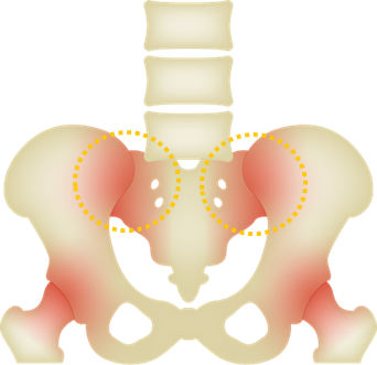
The sacroiliac joint is located in the area that connects the upper and lower body, and is constantly subjected to high pressure and has very little mobility (only a few millimeters), making it a very structurally stable joint.
It is also a pain-sensitive area due to the abundance of muscles and ligaments surrounding it.
The sacroiliac joint is an important part of the body that supports the weight of the upper body, interlinks with the lower body, and is responsible for the balance of the body.
When the joint is slightly misaligned due to work at the middle back, external shock, or childbirth, pain can occur on one or both sides.
Most common age of onset: Teens to 30s (women who have experienced growth spurts, pregnancy/childbirth)
Commonly reported pain site: Near the superior (upper) posterior (back) iliac spine
The pain is often located in the lower back or buttocks and can sometimes be mistaken for other low back pain conditions.
People who commonly develop sacroiliac disorders:
- 1. Work that involves repetitive one-legged loading or repetitive weight shifting (e.g., standing line work, cooks, etc.)
- 2. Women who have experienced pregnancy or childbirth
- 3. Sports such as soccer or softball, where a large force is exerted on a single leg.
- 4. Work that requires a lot of heavy lifting
Sacroiliac arthritis is not rare, and is a common cause of back pain not only during pregnancy and childbirth, but also in men and women of all ages.
Main symptoms of sacroiliac joint disorder
Major Symptoms of Sacroiliac Joint Disorders:
- i. Localized low back pain identified in "Fortin's Sign", a test method in which the patient is asked to point at the painful area with his/her index finger. A positive test is when the patient twice identifies the painful region as within 1 cm of the superior posterior iliac spine.
- ii. Pain and numbness in the buttocks (along the sacroiliac joint surface) and groin
- iii. Pain and numbness in the lower extremities
- iv. Pain and numbness in the lower limbs (often on the outside)
In recent years, some acute back pain (like back cricks) are thought to be caused by a sprain of the sacroiliac joint, and continued twisting of the sacroiliac joint can lead to chronic back pain.
Everyday activities that often causes pain in patients:
- i. Sitting in a chair for long periods of time
- ii. Sleeping on the side that is affected
- iii. Putting weight on one leg
- iv. Walking and running with weight in the toes
- v. Pain at the start of walking but gradually becomes easier (intermittent claudication-like symptoms)
Symptoms in the lumbar buttocks and lower extremities are similar to neurological symptoms such as disc herniation. Sciatic nerve symptoms may also occur and may be complicated by lumbar spine disease.
If you think you might experience of any of these symptoms, we recommend that you have them checked out instead of putting them off.
Causes of sacroiliac joint disorder
Sacroiliac Joint Disorder can be caused by:
- Inflammation due to minor damage to the sacroiliac joint surface caused by repetitive movements such as working in a slouched posture (counter-rotation), twisting the waist, etc.
- Trauma such as a fall from a high place or a traffic accident
- Inflammation of ligaments caused by chronic stress on the surrounding ligaments due to instability caused by misalignment of the sacroiliac joints.
- Inflammation of the sacroiliac joint and surrounding ligaments due to instability of the pelvis caused by weakness of the muscles around the pelvis
- Inflammation caused by damage to ligaments around the sacroiliac joint due to childbirth.
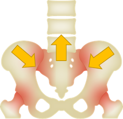
(Nutation)
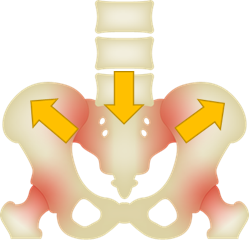
(counternutation)
Tightening Position (Nutation)
A position in which the joints have a lot of contact surfaces and the ligaments and joint capsules are under tension.
A position in which the joints can be stabilized without the use of muscles.
Example: Standing straight
Loosening position (Counter-nutration)
A position in which there is little contact between the joint surfaces and the ligaments and joint capsules are so loose that
A position in which muscles must be used to stabilize the joint
Example: Bending over
Although the above causes have been generally pointed out as conventional causes, recent studies have shown that intervertebral disc degeneration is actually one of the most important factors in sacroiliac joint dysfunction.
(Reference: Unexpected Sex differences in the Relationship of Sacroiliac Joint and Lumbar Spine Degeneration, 2022)
At our clinic, we also believe that there are cases in which the instability of the intervertebral discs are caused by disc degeneration due to a distorted posture and unbalanced muscle stress, which in turn causes inflammation and pain due to the continuous stress on the sacroiliac joint, which serves as the connection between the upper and lower body. Therefore, if MRI images confirm disc degeneration, it is crucial to treat the root cause of disc degeneration as well.
What are the effective treatments for sacroiliac joint disorder?
In many cases, if the MRI scan does not show disc degeneration or spinal canal stenosis, the patient will get better with conservative treatment (an MRI scan is necessary for a more accurate diagnosis because what can be seen on an x-ray alone is different from an MRI). Conversely, if an MRI scan confirms disc degeneration or spinal canal stenosis, it is necessary to treat the disc degeneration or spinal canal stenosis.
Conventional surgical procedures often involve surgical methods to reduce nerve compression. In the case of spinal canal stenosis, spinal fusion is a method that remove muscles and ligaments to fix unstable bones, or an endoscope is used to shave off some lumbar vertebrae and enlarged ligaments in order to reduce nerve compression. However, sometimes the inflammation does not subside and does not improve after surgery, or the pain recurs. Surgery is not a fundamental treatment as the cause of the pain may not only be related to the spine or intervertebral disc, but also other factors such as body constitution, muscles, and ligaments.
When herniated discs or spinal stenosis occur, the pressure on the nerves and the worsening of symptoms are attributed to disc deformity.
Therefore, our clinic believes that if the deformation of the intervertebral disc can be controlled, bone deformation, thickening of the ligaments, and other inflammation related to nerve compression can be prevented.
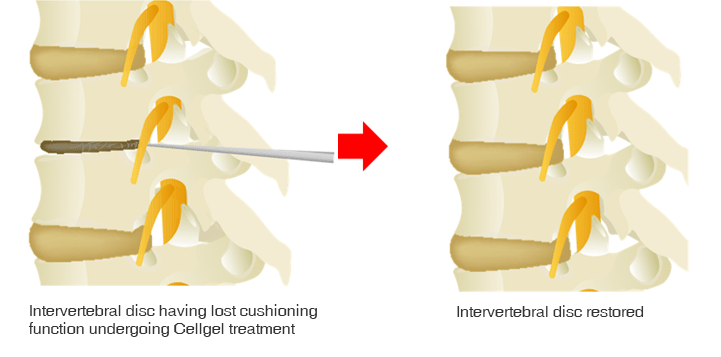
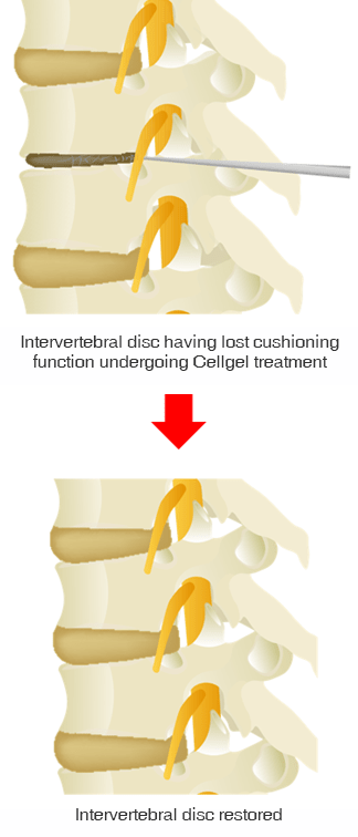
Our disc repair treatment using the Cellgel method restores disc function by repairing the torn annulus fribrosus and replenishing the water content of the nucleus pulposus inside.
The treatment can be performed as a one-day procedure. Local anesthesia is given to the lower back, a needle is inserted into the disc, and a chemical solution is injected.
It takes about 25 minutes and patients can go home after resting for about an hour.
Conventional Surgery
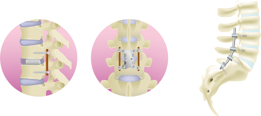
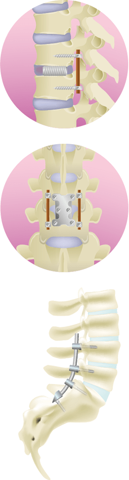
The removal of the bone and part of the disc that is compressing the spinal canal relieves pressure on the spinal canal, but the spine becomes unstable, so an artificial disc is inserted and the bone is secured with a metal device called a bolt and rod.
Cellgel Method
Since many of our patients come from far away,
we are happy to give free consultations to see which treatment is best for you
We'll diagnose your MRI images for free!
Treatment at ILC
There are four main treatments at our clinic.
As mentioned above, the root cause of each back pain disease is "inflammation and deformation due to aging of the intervertebral discs." Although the four treatment methods have different features and advantages, we would like to first introduce the "Cellgel method," which is the only one capable of intervertebral disc repair.
The Cellgel Method
About Cellgel
The Cellgel method is an advanced treatment for low back pain that has been implemented in more than 54 countries worldwide, predominantly in Europe.
As with other treatments, it does not decrease the disc volume, and since the drug remains in the disc as a gel-like implant after treatment, the disc can be preserved. Recent studies have also shown that the volume of the disc increases after treatment.*1
It is also thought that by repairing the disc and preventing leakage of the Nucleus Pulposus, the disc itself will restore its normal function with its own regenerative ability (*2).
It is particularly recommended for patients who are concerned about the risk of conventional disc surgery (MED, PELD, etc.).
*1 Source: European Journal of Radiology 109 (2018) 101–107 , Efficiency of an ethyl alcohol gel in symptomatic disc hernation
*2 Source: International Journal of Spine Surgery Vol. 15 Appendix 1 from regenerative treatment of disc degeneration
"Very good or good results were obtained in 202 (91.4%) of the 221 patients in group A. Of the 44 patients in group B, 37 patients (84%) presented very good or good results and in 9 (82%) of the 11 patients of group C, we obtained similar results. There was no allergic complication in any of our patients. Long-term follow-up magnetic resonance showed a dramatic reduction in hernia volume."
- Dr. J. Theron, one of the world's leading experts in the treatment of Celgel method, from his research article "Percutaneous Treatment of Lumbar Intervertebral Disk Hernias With Radiopaque Gelified Ethanol - A preliminary study."
Merits of Cellgel
- Possibility to repair intervertebral discs where unable to using surgical or laser treatment
- As the disc is repaired, the disc itself regains its original normal function through its own regenerative ability
- The Cellgel method uses local anesthesia over general anesthesia used in conventional surgery, making it less burdensome on the body
- Treatment is done with a thin needle, so the wound is minimal and recovery is rapid
- Treatment is very short, allowing for outpatient day treatment
- Safe, modern treatment proven in more than 54 countries around the world
Flow Cellgel Treatment Process
-
01 Examination

MRI and X-rays will be taken, followed by a doctor's diagnosis.
If the diagnosis proves the Cellgel method suitable, treatment can begin that same afternoon. -
02 Before

TreatmentAfter entering the treatment room and administering local anesthesia to the lower back, a needle is inserted into the intervertebral disc identified in the examination.
A contrast scan is performed to confirm the location of the damaged disc. -
03 Treatment

The Cellgel drug is administered to the damaged area while confirming the location of the disc with a fluoroscopy device.
Once the drug has been absorbed, the needle is removed and the bleeding is stopped.
-
04 Returning

HomeAfter the treatment, the patient rests in a private room for about an hour, and then allowed to go home after the post-treatment examination.
*Resting time depends on the patient's symptoms and condition.
Price Cost of Treatment for the Cellgel Method
| Number of Discs Treated |
1 disc | 2 discs | 3 discs | 4 discs | 5 discs |
|---|---|---|---|---|---|
| Treatment Cost | 1,320,000 JPY | 1,430,000 JPY | 1,540,000 JPY | 1,650,000 JPY | 1,760,000 JPY |
Swipe left/righ
* The cost of treatment is indicated including taxes.
*If treatment is performed at our clinic, all examination/diagnosis costs and test costs such as MRI are included in the above costs.
*This treatment method is not covered by Japanese health insurance, so you will have to pay for the entire treatment yourself.
*Payment by credit card (VISA, MasterCard, JCB, American Express, Diners, Discover) is also possible.
*If you would like rehabilitation (low back pain specialized rehabilitation), it is also possible to pay by bank transfer.
*If you live and work in Japan, you can receive a tax refund by filing your final medical expense return.
FAQ FAQ about sacroiliac joint disorder and treatment
-
QWhat's the difference in the reccurence rate between Cellgel and conventional surgery?
-
A
Surgical procedures, which began in the 1960s, aimed to remove and sometimes fix deformed bones and herniated tissue. However, new bone damage caused by screws and the lack of fundamental treatment (repair of the intervertebral disc annulus fibrosus) resulted in the appearance of additional back pain and recurrence rates.
Therefore, in the 1980s, the need for fundamental treatment increased, and the intervertebral disc treatment (Cellgel) that we provide at our clinic began. Cellgel, in particular, have been proven to repair and regenerate the intervertebral disc, resulting in not only improvement of symptoms, but also an extremely low recurrence rate.
-
QHow is the disc to be treated determined?
-
AThe doctor will spend about 30 minutes with the patient while looking at the MRI and X-ray images to determine the cause of the condition, determine the treatment area, and explain the corresponding treatment method to the patient. MRI images show the main factors that contribute to back pain (disc, nerve, ligament, and joint), such as changes in shape, pressure on the nerve, presence or absence of damage, and inflammation. The x-ray image mainly confirms the condition of the bones.
-
QWhy is the Cellgel method applicable to cases where surgery showed no improvement?
-
ADuring surgery for disc herniation, the prolapsed herniation is removed, but the damaged disc remains intact. As a result, fresh nucleus pulposus components leak from the damaged disc and inflammation persists. On the other hand, our treatment can treat the disc injury itself, thus suppressing inflammation by reducing the fresh leakage of nucleus pulposus components.
-
QMy back pain may temporarily worsen after treatment. How long will it last?
-
AThe reason for the lower back pain is that spinal ligaments that have been compressed are stretched; the lower back pain lasts for about two weeks and then subsides.
-
QWhat level of exercise can I perform after treatment?
-
AGeneral recreational exercise and sports can be done without any problem. For professional athletes, the possibility of new disc damage is high and should be discussed in consultation with a doctor.
-
QPlease tell me about what I should be cautious about after treatment.
-
ADuring the first few days after treatment, the patient should rest and avoid prolonged sitting, heavy lifting, twisting, bending, or strenuous exercise. Patients should be able to perform routine tasks one week after treatment and should be able to perform light exercise by the second week. Weight training is allowed after 3 months.
-
QIs the treatment covered by the Japanese national health insurance?
-
ANo, it is not covered by the national insurance. If you have life insurance, you may be eligible. Please feel free to consult our staff regarding this matter.
-
QHow long must I wait to be able to walk after treatment?
-
AIt is an outpatient/day treatment, so you can walk about an hour after the surgery.
-
QHow many doctor visits do I need to make before treatment?
-
AWe require an MRI to be taken within three months for remote diagnosis, either with our affiliated clinic or from data sent by you. You can receive treatment on the same day after phyiscal diagnosis. You may also request only the diagnosis and come back only for the treatment at a later date.
-
QCan I fly immediately after the treatment?
-
AIf there are no problems after the treatment, you may fly. If there is any pain after the treatment, flying may not be recommended depending on the doctor's evaluation.
-
QWhat should I be cautious of when I have a sacroiliac joint disorder?
-
AIf you are diagnosed with sacroiliac joint disorder, the first step is to prevent further disc and bone degeneration.
If you perform a lot of activities or tasks that place particular stress on your lower back, reduce the frequency of such activities , and listen carefully to your doctor's advice to determine what to do next.
-
QCan sacroiliac joint disorder be cured without surgery?
-
A
If there are no other complications such as disc herniation or spinal canal stenosis, conservative treatment will probably be sufficient in many cases.
However, it has recently been found that sacroiliac joint disorders are sometimes associated with disc degeneration, so in order to resolve the root cause of the problem, an MRI scan of not only the sacroiliac joint but also the hip bones should be performed for a thorough diagnosis. If disc degeneration is found, corresponding treatment will be necessary.Our Cellgel method is a minimally invasive treatment that can be performed in a day, and requires only an injection under local anesthesia, so we recommend it to those who are anxious about surgery.
-
QCan I continue working if I have a sacroiliac joint disorder?
-
A
It is possible to continue, depending on the severity of the symptoms.
However, if your job involves a lot of desk work or heavy labor, you need to refrain from such work as much as possible, as it is hard on the lower back.
If it is difficult to change your work environment, consider improving your physical condition, posture, and muscle strength. If these methods improve the condition, we recommend that you continue to use them.If there is no improvement, we recommend that you consult a doctor and receive a diagnosis.
-
QWon't I make the sacroiliac joint disorder worse by putting pressure on my body during rehabilitation?
-
ATemporary strain on the sacroiliac joint may be placed during physical evaluation, but when performing rehabilitation exercises, the movements are performed in a way that does not place a burden on the joint.
-
QCan I still do rehabilitation if I am in pain?
-
AThis is not a problem as rehabilitation is performed to alleviate pain and other symptoms.
Pain may appear when you move your body to check its condition, but when you exercise, you will do so in a way that does not cause symptoms.
ILC Rehabilitation X Sacroiliac Joint Disorder Treatment
Although the Cellgel method can repair and regenerate the disc itself, and other treatment methods we offer can fundamentally heal the disc; it is possible that the cause of back pain is not only the disc, but also muscles, joints, and ligaments that are affected by the disc.
In cooperation with our rehabilitation specialists, we recommend treating back pain caused by muscles, joints, and ligaments together.
We also offer a short-term intensive program for patients who live far away or overseas.
-
ILC's Low Back Pain Treatment
Outpatient Treatment
Improve back pain disorders/
symptoms in relation to the nerves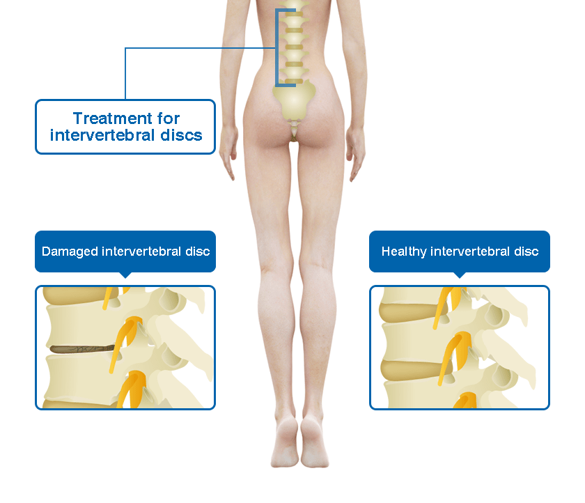
-
ILC's Rehabilitation Program
Specialized Low Back Pain Rehabilitation
Improve back pain disorders/symptoms in
relation to the joints, muscles, ligaments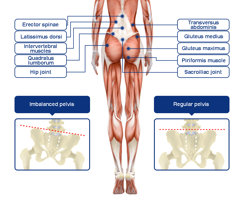
-
01
ILC's Specialized Back Pain Rehabilitation
Our Rehabilitation Approach to Sacroiliac Joint Disorder
The frequency of sacroiliac joint disorders is estimated to be about 25% of patients with low back pain, and data indicates that 39% of these disorders are associated with spinal disorders, making them statistically more prevalent. (Reference: Recognizing specific characteristics of nonspecific low back pain URL:
https://pubmed.ncbi.nlm.nih.gov/2951048/)Sacroiliac joint disorders can be divided into two main categories: those that occur in the sacroiliac joint itself and those that are caused by the ligaments and muscles surrounding the sacroiliac joint.
When the sacroiliac joint itself is affected, it is said to be more mobile than normal. The muscles and ligaments may not be able to stabilize the sacroiliac joint, and intervention with exercise therapy may be required.If the ligaments and muscles surrounding the sacroiliac joint are causing problems, the muscles may not be working properly or may be overworked due to the movements and postures of daily life. It is important to find out what kind of posture or movement is causing the stress.
In addition, if the stress is prolonged and the inflammation is chronic, the localized chronic inflammation will not improve easily, even if the stress is reduced by exercise, because of the sensitivity to pain. In such cases, acupuncture and moxibustion therapy and diet therapy are used in combination to alleviate the inflammation as soon as possible.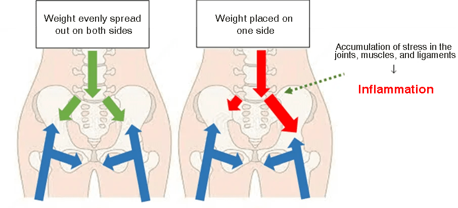
-
02
ILC's Specialized Back Pain Rehabilitation
How to Evaluate Sacroiliac Joint Disorder
Based on the imaging findings, a consultation is held with the doctor to determine what kind of stress is occurring in the sacroiliac joint.
Using these findings as a guide, we evaluate aggravating and mitigating factors of symptoms in daily life postures and movements by slowly moving the body in practical settings.
Aggravating and mitigating factors are defined as movements that should not be performed, and mitigating factors are symptoms that can be alleviated by doing certain body movements.
These factors are comprehensively evaluated with muscle strength and stiffness to find out why the sacroiliac joint disorder is occurring and why pain symptoms are appearing.
If there is any abnormal posture or movement here, we will evaluate the physical factors such as muscle strength and range of motion while slowly moving the body to find out why the body is being used in such a way. -
03
ILC's Specialized Back Pain Rehabilitation
Rehabilitation Treatment for Sacroiliac Joint Disorder
Rehabilitation involves confirming where the pain is located and what is causing the pain.
This confirmation is extremely important to identify the cause, where the strain is likely to be exerted, and what is triggering the pain.
The pelvis is then moved in the direction of pain relief. The pelvis is then repositioned, and the pelvic muscles are strengthened and the hip joints and back are stretched to maintain this position. By correcting the pain-causing movements, we aim to reduce the pain and prevent recurrence.to find out why lumbar instability has developed and pain symptoms have appeared.Treatment Methods:
- Manual therapy (improving range of motion, muscle mobility, stretching, etc.)
- Exercise therapy (acquisition of spinal column support muscles, flexibility and strength of gluteal and adductor muscle groups)
- Acupuncture (improve muscle mobility, reduce friction around joints, alleviate pain)
- Guidance on daily activities to reduce increased stress on the sacroiliac joint
- Guidance on self-exercises that can be done at home
-
04
ILC's Specialized Back Pain Rehabilitation
Treatment Period for Sacroiliac Joint Disorder
Even if the cause is the same sacroiliac joint, depending on the condition of the joint and the length of time the symptoms have been present, the duration of treatment may vary from one or two treatment sessions to years.
We aim to achieve 100% effectiveness of treatment while incorporating the latest technology, so we spend at least 30 minutes with each patient to carefully and meticulously examine the patient's concerns in order to make a proper diagnosis and propose the most suitable treatment methods.
We are certain to find a way to alleviate or eliminate the pain and numbness you are experiencing, so please do not give up and give us a call instead!
- Tadaaki Minowa, Clinic Director