

Column How to Interpret MRI Images (Part 3) | ILC International Low Back Pain Clinic (Tokyo) 🔍
January 14, 2023
This analysis continues our interpretation of MRI images, focusing on the combination of sagittal and axial views to pinpoint spinal compression.
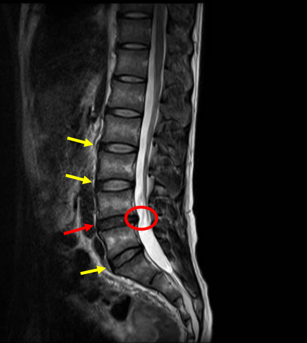
Sagittal T2-Weighted View
Observation: You can see that the disc marked in red (L4/5) is darker (in black) than the disc marked in yellow.
Interpretation: This darker disc indicates disc degeneration caused by the loss of water content in the disc.
Focusing on the red circle, you can see the disc material protruding backward and compressing the spinal canal.
Let’s examine this compression in more detail using the axial (cross-sectional) view.
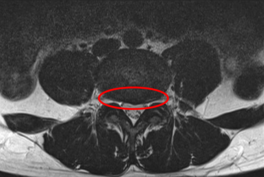
Axial T2-Weighted View
This image is a cross-sectional view corresponding to the sagittal view shown earlier, confirming spinal canal compression.
The red circle highlights the area of compression.
Interpretation: The dark (in black) area within the circle represents the degenerated disc material (a bulge or protrusion). This material is clearly seen compressing the spinal canal and both nerve roots as they exit the central canal.
These findings are typically caused by disc bulging (protrusion).
Based on imaging findings, patient interview, and physical examination, an appropriate treatment plan is determined in consultation with the patient.
Author of this article: Tadaaki Minowa, Administrative Director at ILC Clinic Tokyo