

Column The proper Way to Look at a MRI Scan of The Cervical Spine
August 09, 2023
From this month, we have started treatment of cervical spine at our clinic. As the number of cervical spine cases treated at our clinic increase, I will also be updating the articles on cervical spine treatment. In the meantime, I would like to explain how to look at a basic MRI scan of the cervical spine.
MRI of the cervical spine
There are seven cervical vertebrae in the human body.
On an MRI, they are counted as follows:
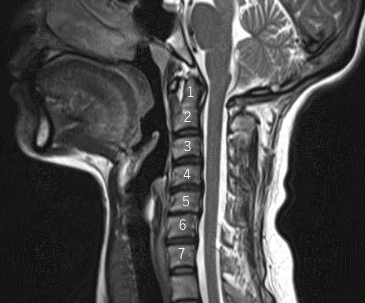
The first and second cervical vertebrae are called the atlas and the axis and are connected to each other like a nut and bolt.
The image below shows the structure of those two vertebrae:
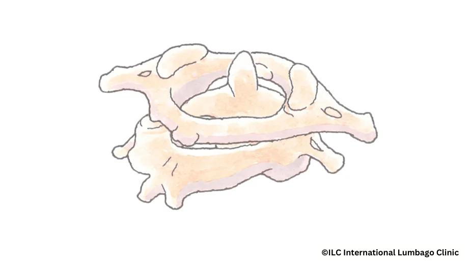
C1 and C2 overlap each other, so there is no intervertebral disc in between.
Therefore, the treatment range of the Cellgel method for intervertebral disc treatment is from C2/3 to C7/T1.
Imaging Anatomy
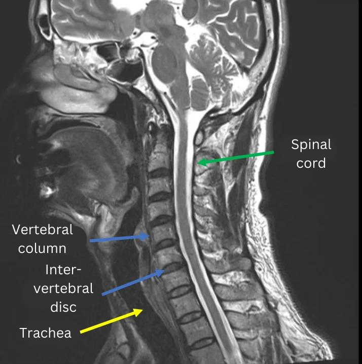
Sagittal section (longitudinal cut image):
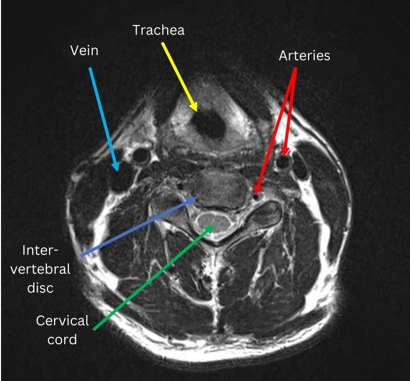
Transverse section (transverse cut):
The structures of the vertebral bodies and intervertebral discs in the cervical area are essentially the same as in the lumbar spine.
However, the size is smaller in the cervical spine, and the intervertebral discs are also smaller than in the lumbar spine.
Also, unlike the lumbar spine, the trachea and multiple blood vessels are in close proximity, and the spinal nerves run behind the intervertebral discs.
This is the basic anatomy of the cervical spine. I will write about findings and names of the conditions in future columns on cervical spine treatments.
If you are currently suffering from neck pain or pain and numbness in your hands and shoulders, please contact us for a consultation.
Phone: 03-6712-3520 (9:00-17:00 except Sunday)
Mail address: click here
If you have an MRI of the cervical spine at hand, please contact us for a free imaging consultation.
*In principle, MRI images should be taken within the last 3 months.
This article was written by a staff member of ILC Tokyo