

Column How to Interpret MRI Images (Part 1) | ILC International Low Back Pain Clinic (Tokyo) Column 🔍
January 7, 2023
This analysis focuses on interpreting basic findings of intervertebral disc health using MRI T2-weighted images.
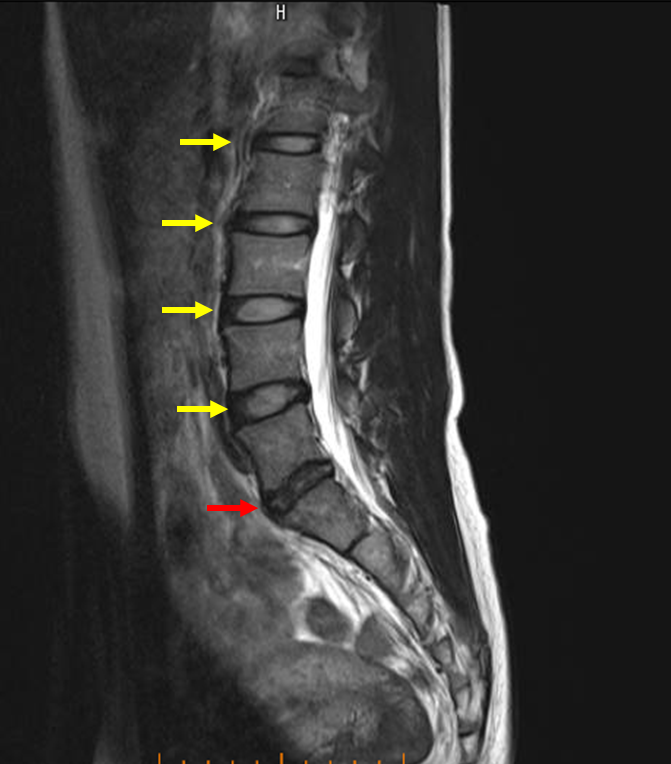
Sagittal T2-Weighted View
Observation: It is clearly visible that the disc marked in red appears darker (or black) than the other discs, such as the one marked in yellow.
Interpretation: This darkening is a key visual sign of disc degeneration. The disc has lost water content, reducing its signal intensity on the T2 image.
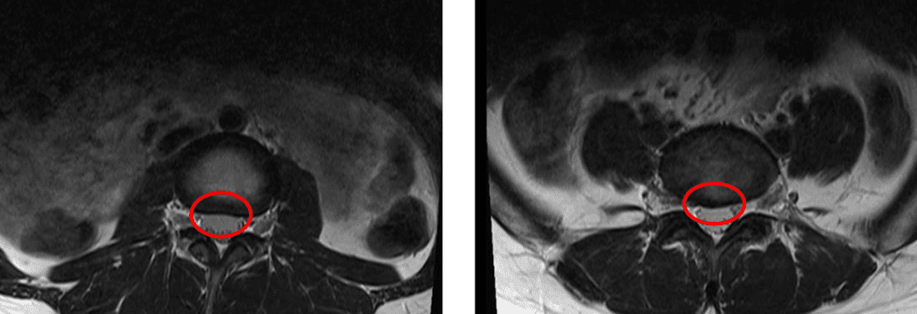
Axial T2-Weighted View
This image provides a cross-sectional view. The image compares a normal disc (left) with an abnormal disc (right).
Observation: By looking closely, you can see that the disc on the right, marked by the red circle, is bulging or protruding and is compressing the spinal canal.
Interpretation: This bulging is caused by the underlying disc degeneration. This type of finding can lead to pain and nerve symptoms.
The final treatment strategy is determined through a comprehensive process that includes an evaluation of the image findings, patient interview, and physical examination, followed by a consultation with the patient.
Author of this article: Tadaaki Minowa, Administrative Director at ILC Clinic Tokyo