

Column How to Interpret MRI Images (Part 2) | ILC International Low Back Pain Clinic (Tokyo) Column 🔍
January 10, 2023
This analysis focuses on interpreting specific MRI views related to disc issues.
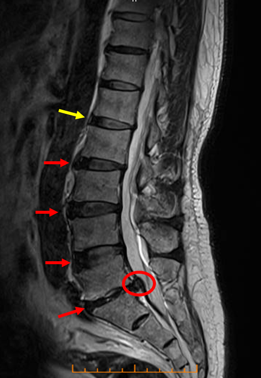
Sagittal T2-Weighted View
Observation: In this image in Sagittal T2-Weighted View, you can see that the disc highlighted in red appears darker compared to the disc highlighted in yellow.
Interpretation: While L1/2 appears relatively normal, levels L2/3, L3/4, L4/5, and L5/S all show signs of disc degeneration.
The disc at the very bottom, L5/S, is visibly compressing the spinal canal.
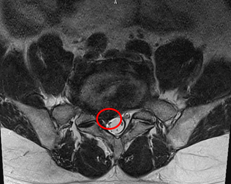
Axial T2-Weighted View
This image provides a cross-sectional view corresponding to the level where spinal canal compression was observed in the sagittal image.
Observation: You can see that the area circled in red appears dark.
Interpretation: This dark area represents the degenerated disc material (a herniation or severe bulge) that is directly compressing the spinal canal.
These imaging findings are consistent with lumbar disc herniation.
Based on imaging findings, patient interview, and physical examination,
we determine the most appropriate treatment plan in consultation with the patient.
Author of this article: Tadaaki Minowa, Administrative Director at ILC Clinic Tokyo